 "
"
Team:CIDEB-UANL Mexico/project capture
From 2014hs.igem.org
DiegoValadez (Talk | contribs) |
DiegoValadez (Talk | contribs) |
||
| Line 331: | Line 331: | ||
</div> | </div> | ||
<div class="container-text"> | <div class="container-text"> | ||
| + | |||
| + | <table width=100%> | ||
| + | <tr> | ||
| + | <td> | ||
| + | <p>In order to desalinize water our project intends to capture sodium ions from saline water using a protein produced from the NhaS gene expression. NhaS is a putative protein from <i>Bacillus firmus</i> that is characterized by its ability to bind and sequestering sodium ions. It “can enhance the Na+ -resistance of antiporter- deficient strains by increasing the availability of Na+ to the integral membrane antiporters on the cytoplasmic side of the membrane and by sequestering Na+ while rate-limiting efflux mechanisms catalyze extrusion of the cation.” (Krulwich & Ivey, 1992)</p> | ||
| + | </td> | ||
| + | <td style="padding-left:12px;"><img width=124 height=113 src="https://static.igem.org/mediawiki/2014hs/e/ec/CapturemoduleCIDEB.jpg"/></td> | ||
| + | </tr></table> | ||
| + | |||
| + | <p>Research by Krulwich and Ivey (1992) supports that in its origin bacteria, NhaS works as a regulation pH homeostasis protein because it makes the cytoplasmic pH more acidic than the external medium, usually basic. The calculated weight of the protein is 7100 Daltons and the final protein product is very basic with a calculated pH of 12.</p> | ||
| + | |||
| + | <p>Basically, NhaS enhance the resistance of bacteria to high saline conditions, regulates pH and captures sodium ions.</p> | ||
| + | |||
| + | <center><p><img width=507 height=221 src="https://static.igem.org/mediawiki/2014hs/6/67/CaptureCIDEB.jpg" | ||
| + | align=center hspace=12 alt="IMG_0317"></p> | ||
| + | |||
| + | <p>Figure 1: Patent US 5346815 A shows extracts of the E. coli EP432 transformed with pGEM (fig. 4A) and pGRVH (fig. 4B). The first one is a control plasmid and the second a plasmid with the NhaS gene. Those are crude extracts that were shown by the effect of putting the bacteria to an SFBI excitation, which is a sodium-sensitive molecule used to measure intracellular Na+. Resuming, it shows in basic draws that the protein is expressed in E. coli and in what quantity according to the excitation level where it is exposed.</p></center> | ||
| + | |||
| + | <p><b>Research on NhaS</b></p> | ||
| + | |||
| + | <p>It is important to be familiarized with what it is being worked with, and since this putative gene has never been used at iGEM before, we did a lot of research on it. </p> | ||
| + | |||
| + | <p>The composition and form of a protein show relevant data about its actions and functions, that is why we investigated NhaS’ predicted type. We found in the modelling tool <a href="http://bmm.cancerresearchuk.org/~3djigsaw/">3D-JIGSAW</a> from Cancer Research UK's site that its possible protein or peptide type would be helix, coil or strand.</p> | ||
<div style="text-align: right;"><a href="https://2014hs.igem.org/Team:CIDEB-UANL_Mexico/project_capture#"><font color="blue">Return to the Top</font></a></p></div> | <div style="text-align: right;"><a href="https://2014hs.igem.org/Team:CIDEB-UANL_Mexico/project_capture#"><font color="blue">Return to the Top</font></a></p></div> | ||
| + | |||
| + | <center><p><img width=507 height=221 src="https://static.igem.org/mediawiki/2014hs/5/54/PPRESULTS.jpg" | ||
| + | align=center hspace=12 alt="IMG_0317"></p> | ||
| + | |||
| + | <p>Figure 2: Interactive 3D-Jigsaw's result that indicates the predicted protein type of NhaS.</p></center> | ||
| + | |||
| + | <p>The previous information was confirmed in Predict Protein site.</p> | ||
| + | |||
| + | <center><p><img width=507 height=221 src="https://static.igem.org/mediawiki/2014hs/9/98/GraphsCIDEB.jpg" | ||
| + | align=center hspace=12 alt="IMG_0317"></p> | ||
| + | |||
| + | <p>Figure 3: Results given by Predict Protein showing the secondary structure composition and solvent accessibility of the putative NhaS gene.</p></center> | ||
| + | |||
| + | <center><p><img width=507 height=221 src="https://static.igem.org/mediawiki/2014hs/5/5b/PPCIDEB.jpg" | ||
| + | align=center hspace=12 alt="IMG_0317"></p> | ||
| + | |||
| + | <p>Figure 4: Results given by Predict Protein showing the predicted precise structure of the NhaS protein. </p></center> | ||
| + | |||
| + | <p>Based on the previous information we conclude that NhaS is most possible to be of the helix type. | ||
| + | Being aware of the secondary structure of proteins is relevant, since hence, the protein folding mechanism can be taken into consideration.</p> | ||
| + | |||
| + | <p>According to Krulwich and Ivey (1992), the location of the protein is in the cytoplasmic side of the membrane, however, when we made our research, we found out that the protein is predicted to be highly non-cytoplasmic (Yeast Resource Center, 2014). We came out with a hypothesis in which NhaS would be located in the inner part of the membrane but on its cytoplasmic side. This would explain both predictions of both sources of information.</p> | ||
| + | |||
| + | <center><p><img width=507 height=221 src="https://static.igem.org/mediawiki/2014hs/3/32/ProteinCIDEB.jpg" | ||
| + | align=center hspace=12 alt="IMG_0317"></p> | ||
| + | |||
| + | <p>Figure 5: Predicted protein overview results from Yeast Resource Center.</p></center> | ||
| + | |||
| + | <p>As a “confirmation” for our hypothesis, Predict Protein site gave us the following result:</p> | ||
| + | |||
| + | <center><p><img width=507 height=221 src="https://static.igem.org/mediawiki/2014hs/2/28/PPCIDEB2.jpg" | ||
| + | align=center hspace=12 alt="IMG_0317"></p> | ||
| + | |||
| + | <p>Figure 6: “Predicted localization for the Bacteria domain: Inner Membrane (GO term ID: 0005886) Prediction confidence 76.”</p></center> | ||
| + | |||
| + | <p>Based on this predicted result and with the previous hypothesis we formulated, the team concluded that the protein would act in the cytoplasmic side of the inner membrane. This information was used for the understanding and explaining of the module, as well as for designing different animations.</p> | ||
| + | |||
| + | <p>Further information about NhaS can be found in its parts registry section.</p> | ||
| + | |||
</div> | </div> | ||
Revision as of 01:13, 13 June 2014
|
|


Capture Module
|
In order to desalinize water our project intends to capture sodium ions from saline water using a protein produced from the NhaS gene expression. NhaS is a putative protein from Bacillus firmus that is characterized by its ability to bind and sequestering sodium ions. It “can enhance the Na+ -resistance of antiporter- deficient strains by increasing the availability of Na+ to the integral membrane antiporters on the cytoplasmic side of the membrane and by sequestering Na+ while rate-limiting efflux mechanisms catalyze extrusion of the cation.” (Krulwich & Ivey, 1992) |
 |
Research by Krulwich and Ivey (1992) supports that in its origin bacteria, NhaS works as a regulation pH homeostasis protein because it makes the cytoplasmic pH more acidic than the external medium, usually basic. The calculated weight of the protein is 7100 Daltons and the final protein product is very basic with a calculated pH of 12.
Basically, NhaS enhance the resistance of bacteria to high saline conditions, regulates pH and captures sodium ions.

Figure 1: Patent US 5346815 A shows extracts of the E. coli EP432 transformed with pGEM (fig. 4A) and pGRVH (fig. 4B). The first one is a control plasmid and the second a plasmid with the NhaS gene. Those are crude extracts that were shown by the effect of putting the bacteria to an SFBI excitation, which is a sodium-sensitive molecule used to measure intracellular Na+. Resuming, it shows in basic draws that the protein is expressed in E. coli and in what quantity according to the excitation level where it is exposed.
Research on NhaS
It is important to be familiarized with what it is being worked with, and since this putative gene has never been used at iGEM before, we did a lot of research on it.
The composition and form of a protein show relevant data about its actions and functions, that is why we investigated NhaS’ predicted type. We found in the modelling tool 3D-JIGSAW from Cancer Research UK's site that its possible protein or peptide type would be helix, coil or strand.

Figure 2: Interactive 3D-Jigsaw's result that indicates the predicted protein type of NhaS.
The previous information was confirmed in Predict Protein site.

Figure 3: Results given by Predict Protein showing the secondary structure composition and solvent accessibility of the putative NhaS gene.

Figure 4: Results given by Predict Protein showing the predicted precise structure of the NhaS protein.
Based on the previous information we conclude that NhaS is most possible to be of the helix type. Being aware of the secondary structure of proteins is relevant, since hence, the protein folding mechanism can be taken into consideration.
According to Krulwich and Ivey (1992), the location of the protein is in the cytoplasmic side of the membrane, however, when we made our research, we found out that the protein is predicted to be highly non-cytoplasmic (Yeast Resource Center, 2014). We came out with a hypothesis in which NhaS would be located in the inner part of the membrane but on its cytoplasmic side. This would explain both predictions of both sources of information.

Figure 5: Predicted protein overview results from Yeast Resource Center.
As a “confirmation” for our hypothesis, Predict Protein site gave us the following result:
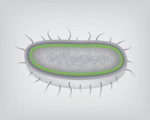
Figure 6: “Predicted localization for the Bacteria domain: Inner Membrane (GO term ID: 0005886) Prediction confidence 76.”
Based on this predicted result and with the previous hypothesis we formulated, the team concluded that the protein would act in the cytoplasmic side of the inner membrane. This information was used for the understanding and explaining of the module, as well as for designing different animations.
Further information about NhaS can be found in its parts registry section.
Av. Lázaro Cárdenas to the
East n/a Col. Mederos, Monterrey, Nuevo León, México.
Pedro de Alba n/a Ciudad Universitaria, San Nicolás de Los Garza, Nuevo León, México.

igem.cidebuanl@gmail.com


iGEM CIDEB UANL 2014. CENTRO DE INVESTIGACIÓN Y DESARROLLO DE EDUCACIÓN BILINGÜE. UANL